
A histopathological analysis of 119 surgically excised loose bodies revealed that the cases could be separated into three categories. Coronal 1A axial 1B 1D and sagittal 1C fat.

Synovial Chondromatosis Springerlink
Pathology of the Popliteus Tendon James Y.

. A tissue is a functional aggregation of similar cells and their intercellular materials that combine to perform common functions. Menisci also known as semi-lunar cartilages referring to their half-moon crescent shape. Taco Geertsma Retired Radiologist Gelderse Vallei Hospital.
We histologically examined 84. Methods We histologically examined 84 loose bodies and 9 related lesions synovial membrane. Crescent-shaped fibrocartilaginous structure that in contrast to articular disks only partly.
493-523 Article A study of loose bodies composed of cartilage or of cartilage and bone. Loose bodies are usually asymptomatic 1. An 84-year-old male presents with lateral knee pain and swelling.
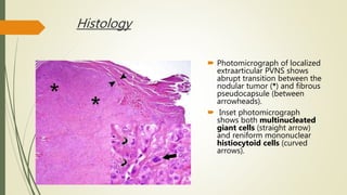
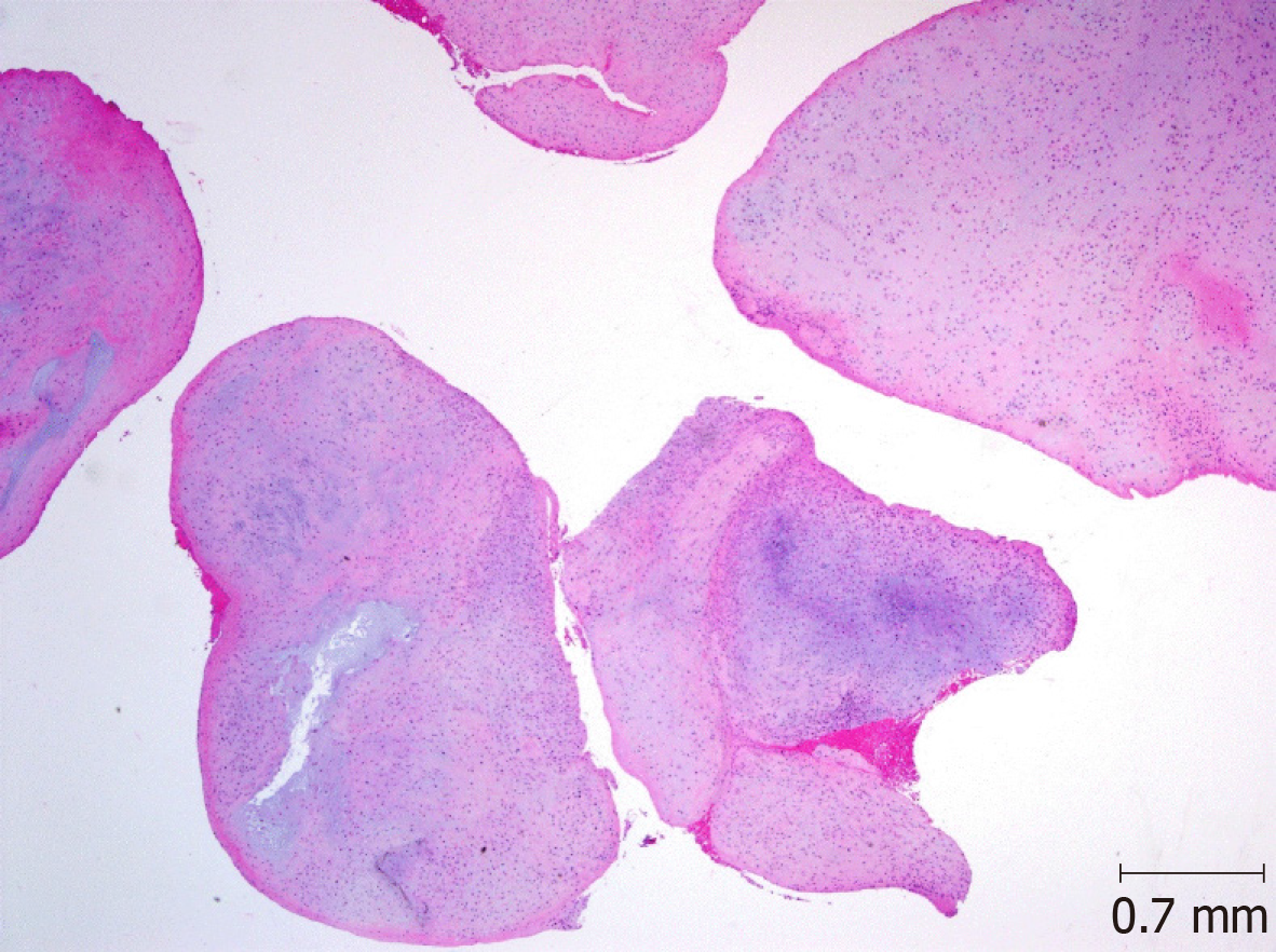
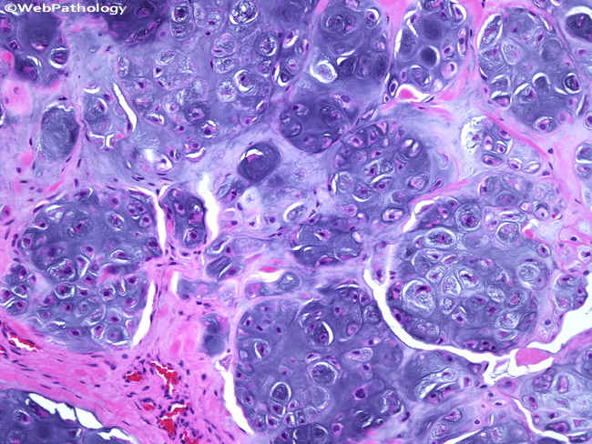
Synovial Chondromatosis is a proliferative disease of the synovium associated with cartilage metaplasia that results in multiple intra-articular loose bodies. Loose bodies in the bicipital groove. Histologically based analyses of the nature and origin of loose bodies occurring in osteoarthrosis have been few and further study is warranted.
General HPB Lower GI Upper GI Vascular BJS British Journal of Surgery Volume 8 Issue 32 p. The loose bodies can vary in size from a few millimeters such as the size of a small pill to a few centimeters the size of a quarter. The condition usually presents.
1 loose bodies due to synovial osteochondromatosis. Loose body and effusion in the biceps tendon sheath in a patient with. Histologically based analyses of the nature and origin of loose bodies occurring in osteoarthrosis have been few and further study is warranted.
An organ is an anatomically discrete structure. The fragments can lead to damage to the articular cartilage. This region is also a loose irregular connective tissue but can be so extensively infiltrated by white blood cells and plasma cells that the supporting fibers and ground substance are.
Loose bodies in the bicipital groove. Methods We histologically examined 84 loose bodies and 9 related lesions synovial membrane nodules surgically. Pathology Peritoneal loose bodies are formed by the torsion and autoamputation of epiploic appendages.
Up to 10 cash back Histologically based analyses of the nature and origin of loose bodies occurring in osteoarthrosis have been few and further study is warranted. 614 Biceps tendon various pathology. Schammel Abstract and Figures Peritoneal loose bodies PLBs have been sparingly documented within the surgical and radiologic literature with 38 cases reported to.
Taco Geertsma is the founder of UltrasoundCasesinfo and a retired radiologist and has worked in the Gelderse Vallei. Histologically based analyses of the nature and origin of loose bodies occurring in osteoarthrosis have been few and further study is warranted. Blood vessel rich - key element proliferation of fibroblasts - key element inflammation - especially lymphocytes plasma cells common -.

Histologic Evaluation Of Osteochondral Loose Bodies And Repaired Tissues After Fixation Arthroscopy
Pvns Synovial Chondromatosis Loose Bodies

Pathology Outlines Histology Joints

Figure 3 Arthroscopic Treatment Of A Case With Concomitant Subacromial And Subdeltoid Synovial Chondromatosis And Labrum Tear

A Loose Bodies Showing Endochondral Ossification In The Hyaline Download Scientific Diagram

Xmlinkhub

Autoamputated Adnexa Presents As A Peritoneal Loose Body Fertility And Sterility

Qiao S Pathology Infarcted Appendix Epiploica Peritoneal Flickr
Synovial Osteochondromatosis Of The Temporomandibular Joint A Case Report

A Large Loose Body Of Around 20x19x6 Cm Extracted From The Knee Joint Download Scientific Diagram

Qiao S Pathology Infarcted Appendix Epiploica Peritoneal Flickr
Webpathology Com A Collection Of Surgical Pathology Images

Synovial Chondromatosis Synovial Osteochondromatosis Surgical Pathology

Macro And Microscopic Findings Of The Loose Body A Extracted Loose Download Scientific Diagram

Gross Finding Of Loose Bodies Several Loose Bodies Shown After Removal Download Scientific Diagram
Histological Examination Of The Loose Bodies Fibrous Connective Tissue Download Scientific Diagram

Qiao S Pathology Infarcted Appendix Epiploica Peritoneal Flickr
Cartilage Proliferation Is Diagnosed During Histopathologic Evaluation Download Scientific Diagram

A Rice Bodies Gross Specimen B Rice Bodies Microscopic Histology Download Scientific Diagram